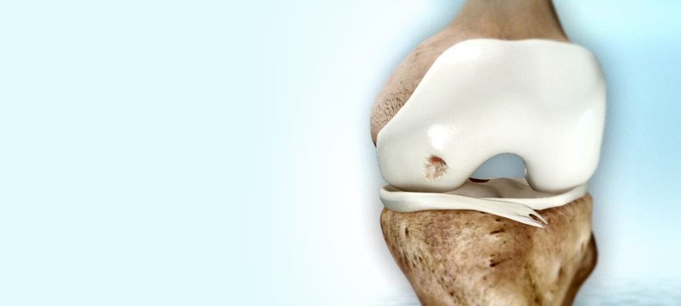
What are joint surface lesions of the knee?
The knee joint consists of two main tissues: articular cartilage and subchondral bone. Together they form the load-bearing system that enables the joint to move freely and without pain. Articular cartilage covers the subchondral knee joint, protecting it, absorbing shock, distributing the load, and facilitating smooth, stable motion.


Joint surface lesions (JSL) involving the articular cartilage and subchondral bone are a common orthopedic problem. JSLs can be superficial, partial-thickness cartilage defects, which do not involve the subchondral bone, or full thickness lesions, which cross the osteochondral junction.
JSLs are very common, and reported in about 20% of all arthroscopic procedures. They are clinically important, as they can be symptomatic and disabling, with pain and/or locking of the joint, and can lead to further cartilage loss and development of OA.
If left untreated, JSLs can lead to secondary osteoarthritis (OA) mainly due to the poor self-healing ability of articular cartilage. Symptomatic chronic full-thickness defects of the knee joint surface may require surgical intervention to relieve symptoms and prevent possible deterioration to OA.


